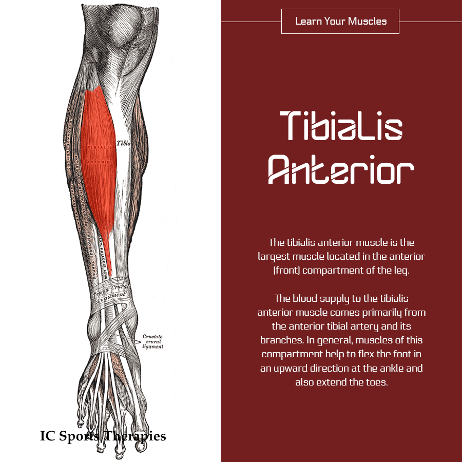
Your Monday Muscle: #9 Tibialis Anterior

Tibialis Anterior

The tibialis anterior muscle is the largest muscle located in the anterior (front) compartment of the leg. The blood supply to the tibialis anterior muscle comes primarily from the anterior tibial artery and its branches. In general, muscles of this compartment help to flex the foot in an upward direction at the ankle and also extend the toes.[1]

The tibialis anterior lies along the outside of the shinbone (tibia). The muscle attaches to the top of the shinbone and descends down the leg, following the outside of the bone. The muscle’s tendon crosses the top of the foot by the inside ankle and connects to two bones (medial cuneiform and first metatarsal) on the bottom of the foot.[2]

What are the pain and symptoms associated with the tibialis anterior muscle?

  • Pain in the big toe
  • Pain in the front of the ankle going up the front of the shin
  • Occasionally there will be swelling of the shin bone
  • Can contribute to shin splints
  • Can be a cause of weak ankles
  • Can contribute to drop foot which can cause tripping and falling [2]

Interesting facts about the tibialis anterior muscle:

  • Trigger points and a tight shortened tibialis anterior can make it difficult to pick up the foot and can contribute to ‘tripping over your own feet’.
  • Pain from trigger points in the tibialis anterior is sometimes diagnosed as gout or turf toe.
  • Is often the primary cause of  “growing” pains in the feet and ankles of children. [2]

Sources:

[1] Tibialis Anterior Healthline

[2] Tibialis Anterior Muscle: Big Toe, Ankle and Shin Pain The Wellness Digest

Previous: Your Monday Muscle: #8 Vastus Lateralis

SaveSave

Your Monday Muscle: #8 Vastus Lateralis

Vastus Lateralis

The vastus lateralis muscle

This muscle is located on the side of the thigh. This muscle is the largest of the quadriceps group (often called quads) which also includes the rectus femoris, the vastus intermedius, and the vastus medialis. Collectively, the quadriceps muscle is the largest in the human body and its purpose is to extend the knee. The specific task of the vastus lateralis muscle is to extend the lower leg and allow the body to rise up from a squatting position.

Trigger points in three of the four quadricep muscles are the kingpins behind many cases of knee pain and dysfunction. The vastus lateralis trigger points refer pain to the outside of the thigh, knee, and upper lower leg. They may also cause the “stuck patella” or “locked knee cap” conditions in which the knee cap fails to track up and down naturally during movements of the knee. While the pain from the other quadriceps trigger points is focused only on the knee joint, the vastus lateralis trigger points may also project pain to the outside of the thigh, which may also be confused with IT Band syndrome.

What are the pain and symptoms associated with the vastus lateralis?

  • Knee pain
  • Pain on the side of the thigh extending down into the front and back of the knee
  • Pain under the buttock extending toward the hip joint
  • Pain occasionally descends into the back of the calf
  • Locked knee
  • Extended walking increases pain in the thigh and knee

Fun Facts about vastus lateralis

  • It is the largest of the quadriceps muscles
  • Everyone has trigger points in the vastus lateralis.
  • ‘Growing pains’ in the knees and hips of children can often be traced to the vastus lateralis

Sources:

  1. Vastus Lateralis. healthline.com.
  2. Vastus Lateralis Trigger Points: The Knee Pain Trigger Points, triggerpointtherapist.com
  3. Vastus Lateralis Muscle: Hip, Thigh, Knee Pain, thewellnessdigest.com

[Previous: Your Monday Muscle: #7 Rectus Femoris ]

Your Monday Muscle: #7 Rectus Femoris

Rectus Femoris

Rectus Femoris.

The rectus femoris muscle is one of the four quadriceps muscles of the human body. The others are the vastus medialis, the vastus intermedius (deep to the rectus femoris), and the vastus lateralis. All four parts of the quadriceps muscle attach to the patella (knee cap) via the quadriceps tendon.

The rectus femoris is situated in the middle of the front of the thigh; it is fusiform in shape, and its superficial fibers are arranged in a bipenniform manner, the deep fibers running straight (rectus) down to the deep aponeurosis. Its functions are to flex the thigh at the hip joint and to extend the leg at the knee joint.[1]

Fun Facts about the Quadriceps and rectus femoris

  • Did you know that the Rectus Femoris is the strongest and leanest muscle in the human body?
  • Did you know that the Rectus Femoris is the only quad that isn’t actually attached to the femur (thigh bone) but actually is attached below the knee and to the pelvis. So it can both straighten the leg and bend the hip!?
  • The Latin full formal name for quadriceps is musculus quadriceps femoris, and the meaning literally translates to “four-headed muscle of the femur”.
  • President Bill Clinton had surgery in 1997 to repair the quadriceps tendon in his right knee, which he tore stumbling on steps.
  • Arnold Shwarzenaeger has just about as many muscle fibers in his quads as you do. They’re just thicker.[2]
  • The quadriceps are used for knee extension, cycling, climbing stairs. Squats or leg extensions on a machine will develop this muscle.[3]

There is a fifth muscle of the quadriceps complex that is often forgotten and rarely taught called articularis genus. In addition, recent cadaver studies have confirmed the presence of a sixth muscle, the tensor vastus intermedius.[4]

Sources:

  1. Sportsmedicine: Rectus Femoris. Sportsmedicine.about.com.
  2. Fun Facts – Rectus Femoris, ihavehugequads.weebly.com
  3. Muscle: Interesting Facts, Names, and Functioninfolific.com
  4. Quadriceps femoris muscle, sussie.org

[Previous: Your Monday Muscle: #6 Brachialis ]

Your Monday Muscle: #6 Brachialis

Brachialis


Brachialis

The brachialis muscle is located in the upper arm.

It lies underneath the biceps muscle. It acts as a structural bridge between the humerus, which is the bone of the upper arm, and the ulna, which is one of the forearm bones.

The muscle is innervated by both the musculoskeletal nerve and the radial nerve.

In some people, the muscle may appear doubled. Also called the brachialis anticus, its primary action is to flex the forearm muscles at the elbow.

[Read more at Healthline]

What movements does the brachialis muscle control?

Bending the elbow

Activities that cause brachialis pain and symptoms:

  • Lifting heavy objects with a bent elbow
  • Picking up children
  • Holding up heavy tools
  • Working at the computer
  • Chin ups
  • Playing the oboe, clarinet, and saxophone

[Read more at the Wellness Digest]


[Previous: Your Monday Muscle: #5 Triceps brachii]

[Next: Your Monday Muscle: #7 Rectus femoris]

Your Monday Muscle: #5 Triceps Brachii

Triceps brachii

Triceps Brachii Muscles – the muscles used to straighten your arm

The Triceps Brachii muscles are located on the back of the humerus and more commonly referred to as the triceps. They derive their name from a Latin phrase meaning “three headed arm muscle”,  due to the fact that the triceps muscles have three muscle heads and therefore have three separate origin attachment points. These three heads are called the Lateral, Medial and Long and they connect the Humerus, Scapula and the >Ulna. The Triceps Brachii muscles are primarily responsible for the extension of the elbow joint (straightening of the arm). They are the largest muscles in the upper arms.

As with the Bicep muscle which has two heads and therefore two origin points, the three-headed Triceps muscle has three different origin points. These origin attachment points are as follows;

1. The Long head arises from the lower part of the Glenoid Cavity which is a shallow depression on the scapula where the head of the Humerus fits. This head is responsible for giving the Triceps Muscle the horse shoe shape.

2.The Lateral head  which arises from the upper half of the outside posterior surface of the humerus.

3.The Medial head which arises from the dorsal and inside posterior of the humerus and can usually only be visible closer to the elbow joint as it is mostly covered by the Lateral and Long heads.

— read more at musclesused.com

Fun Facts

Here are some fun facts about triceps muscles that you need to know.

  1. The Triceps muscle makes up 2/3 of the muscle in your arm.
  2. The average triceps muscle is twice as big as the biceps muscle.
  3. The Triceps muscle has 3 separate heads.
  4. The Triceps muscle is mostly responsible for straightening your arm.
  5. The Triceps muscle instigates shoulder and elbow rotation.
  6. More than 90% of a horse’s total muscle weight is located at their triceps.
  7. In many mammals such as dogs, they have a fourth head called the accesory head. It is located between the lateral and medial heads.
  8. The triceps brachii is a contributor to both tennis elbow and golfers elbow.
  9. It and the serratus anterior are known as the boxer’s muscles because these muscles deliver the straight-arm punch.

— sources:


Previous: Your Monday Muscle: #4 Biceps Brachii

Next:

Your Monday Muscle: #4 Biceps Brachii

Biceps brachii

The biceps brachii

The biceps brachii, sometimes known simply as the biceps, is a skeletal muscle that is involved in the movement of the elbow and shoulder.

It is a double-headed muscle, meaning that it has two points of origin or “heads” in the shoulder area. The short head of each biceps brachii originates at the top of the scapula (at the coracoid process). The long head originates just above the shoulder joint (at the supraglenoid tubercle). Both heads are joined at the elbow.

The biceps brachii is a bi-articular muscle, which means that it helps control the motion of two different joints, the shoulder and the elbow.

The function of the biceps at the elbow is essential to the function of the forearm in lifting. The function of the biceps brachii at the shoulder is less pronounced, playing minor roles in moving the arms forward, upward, and sideways.

Although it is generally considered to be doubled headed, the biceps brachii is one of the most variable muscles in the human body. It is common for human biceps to have a third head originating at the humerus. As many as seven heads have been reported.

— Biceps Brachii Origin, Function & Anatomy,  Healthline


Previous: Your Monday Muscle: #3 Pectoralis Major

Next: Your Monday Muscle: #5 Triceps Brachii

Your Monday Muscle: #3 Pectoralis Major

Pectoralis Major

The pectoralis major

The pectoralis major muscle is a large muscle in the upper chest1, fanning across the chest from the shoulder to the breastbone. The two pectoralis major muscles, commonly referred to as the “pecs,” are the muscles that create the bulk of the chest.

A developed pectoralis major is most evident in males, as the breasts of a female typically hide the pectoral muscles.

A second pectoral muscle, the pectoralis minor, lies beneath the pectoralis major.

The pectorals are predominantly used to control the movement of the arm, with the contractions of the pectoralis major pulling on the humerus to create lateral, vertical, or rotational motion.

The pectorals also play a part in deep inhalation, pulling the ribcage to create room for the lungs to expand.

Six separate sets of muscle fiber have been identified within the pectoralis major muscle, allowing portions of the muscle to be moved independently by the nervous system.

Injuries

Injuries to the pectoralis major can occur during weightlifting, as well as other bodybuilding exercises that place excessive strain on the shoulders and chest.

Symptoms of a pec major sprain will include a sudden sharp pain at the front of the upper arm near the shoulder where the pec major tendon attaches. There is likely to be rapid swelling of the front of the shoulder and upper arm. Tests which will reproduce pain and help confirm the diagnosis include getting the patient to pull their arm across the front of the chest or rotate it inwards against resistance. A visible gap or lump in the muscle may appear.

Previous: Your Monday Muscle: #2 the Deltoid

Next: Your Monday Muscle: #4 Biceps Brachii

Sources

[1] The Pectoralis Major muscle, healthline.com

Your Monday Muscle: #2 the Deltoid

Deltoid

The Deltoid

The deltoid muscle is a rounded, triangular muscle located on the uppermost part of the arm and the top of the shoulder. It is named after the Greek letter delta, which is shaped like an equilateral triangle.

The deltoid is attached by tendons to the skeleton at the clavicle (collarbone), scapula (shoulder blade), and humerus (upper arm bone). The deltoid is widest at the top of the shoulder and narrows to its apex as it travels down the arm.

Contraction of the deltoid muscle results in a wide range of movement of the arm at the shoulder due to its location and the wide separation of its muscle fibres… (Read more at InnerBody.com)

The deltoid muscle has three parts; the front or anterior, middle, and back or posterior.

The muscle lifts the arm up sideways. The front part helps to lift the arm up forwards, known as shoulder flexion and the back part helps to lift the arm up backwards, known as shoulder extension.


Previous: Your Monday Muscle: #1 the Trapezius

Next: Your Monday Muscle: #3 Pectoralis Major

Your Monday Muscle: #1 the Trapezius

Trapezius

The trapezius

The trapezius is one of the major muscles of the back and is responsible for moving, rotating, and stabilizing the scapula (shoulder blade) and extending the head at the neck. It is a wide, flat, superficial muscle that covers most of the upper back and the back of the neck. Like most other muscles, there are two trapezius muscles – a left and a right trapezius – that are symmetrical and meet at the vertebral column

The trapezius starts at the occipital bone (at the back of the head) and the spinous processes of the cervical and thoracic vertebrae, then extends across the neck and back to insert via tendons on the clavicle (collar bone), acromion (the summit of the shoulder), and spine of the scapula. The name trapezius is given to this muscle due to its roughly trapezoidal shape. The trapezius can be divided into three bands of muscle fibers that have distinct structures and functions within the muscle: Upper Trapezius, Middle Trapezius and Lower Trapezius.

Read more at InnerBody.com

Your Monday Muscle: #2 the Deltoid