Your Monday Muscle: #3 Pectoralis Major

Pectoralis Major

The pectoralis major

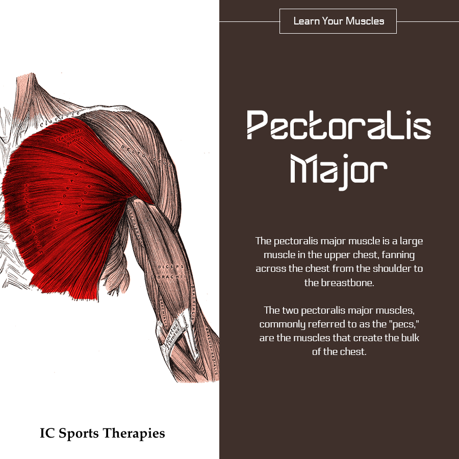
The pectoralis major muscle is a large muscle in the upper chest1, fanning across the chest from the shoulder to the breastbone. The two pectoralis major muscles, commonly referred to as the “pecs,” are the muscles that create the bulk of the chest.

A developed pectoralis major is most evident in males, as the breasts of a female typically hide the pectoral muscles.

A second pectoral muscle, the pectoralis minor, lies beneath the pectoralis major.

The pectorals are predominantly used to control the movement of the arm, with the contractions of the pectoralis major pulling on the humerus to create lateral, vertical, or rotational motion.

The pectorals also play a part in deep inhalation, pulling the ribcage to create room for the lungs to expand.

Six separate sets of muscle fiber have been identified within the pectoralis major muscle, allowing portions of the muscle to be moved independently by the nervous system.

Injuries

Injuries to the pectoralis major can occur during weightlifting, as well as other bodybuilding exercises that place excessive strain on the shoulders and chest.

Symptoms of a pec major sprain will include a sudden sharp pain at the front of the upper arm near the shoulder where the pec major tendon attaches. There is likely to be rapid swelling of the front of the shoulder and upper arm. Tests which will reproduce pain and help confirm the diagnosis include getting the patient to pull their arm across the front of the chest or rotate it inwards against resistance. A visible gap or lump in the muscle may appear.

Previous: Your Monday Muscle: #2 the Deltoid

Next: Your Monday Muscle: #4 Biceps Brachii

Sources

[1] The Pectoralis Major muscle, healthline.com

Friday Fitness Fact #1: The more muscle mass you have…

Muscle Mass

Metabolism: Converting food into energy

Metabolism is the process by which your body converts what you eat and drink into energy. During this complex biochemical process, calories in food and beverages are combined with oxygen to release the energy your body needs to function.

Even when you’re at rest, your body needs energy for all its “hidden” functions, such as breathing, circulating blood, adjusting hormone levels, and growing and repairing cells.

The number of calories your body uses to carry out these basic functions is known as your basal metabolic rate — what you might call metabolism. Several factors determine your individual basal metabolic rate, including:

  • Your body size and composition. The bodies of people who are larger or have more muscle burn more calories, even at rest.
  • Your sex. Men usually have less body fat and more muscle than do women of the same age and weight, burning more calories.
  • Your age. As you get older, the amount of muscle tends to decrease and fat accounts for more of your weight, slowing down calorie burning.

Energy needs for your body’s basic functions stay fairly consistent and aren’t easily changed. Your basal metabolic rate accounts for about 70 percent of the calories you burn every day.

Read more at the Mayo Clinic…


Next: Friday Fitness Fact #2: Sit-ups

Wednesday Wonder #10: 200 million litres of blood

Blood

36 Interesting Facts about the Human Heart

  1. The average adult heart beats 72 times a minute; 100,000 times a day; 3,600,000 times a year; and 2.5 billion times during a lifetime.[5]
  2. Though weighing only 11 ounces on average, a healthy heart pumps 2,000 gallons of blood through 60,000 miles of blood vessels each day.[3]
  3. A kitchen faucet would need to be turned on all the way for at least 45 years to equal the amount of blood pumped by the heart in an average lifetime.[1]
  4. The volume of blood pumped by the heart can vary over a wide range, from five to 30 litres per minute.[6]
  5. Every day, the heart creates enough energy to drive a truck 20 miles. In a lifetime, that is equivalent to driving to the moon and back.[1]
  6. Because the heart has its own electrical impulse, it can continue to beat even when separated from the body, as long as it has an adequate supply of oxygen.[3]
  7. French physician Rene Laennec (1781-1826) invented the stethoscope when he felt it was inappropriate to place his ear on his large-bosomed female patients’ chests.[5]
  8. The foetal heart rate is approximately twice as fast as an adult’s, at about 150 beats per minute. By the time a foetus is 12 weeks old, its heart pumps an amazing 60 pints of blood a day.[7]
  9. The heart pumps blood to almost all of the body’s 75 trillion cells. Only the corneas receive no blood supply.[3]
  10. During an average lifetime, the heart will pump nearly 1.5 million barrels of blood—enough to fill 200 train tank cars.[1]
  11. Five percent of blood supplies the heart, 15-20% goes to the brain and central nervous system, and 22% goes to the kidneys.[1]
  12. The “thump-thump” of a heartbeat is the sound made by the four valves of the heart closing.[1]
  13. The heart does the most physical work of any muscle during a lifetime. The power output of the heart ranges from 1-5 watts. While the quadriceps can produce 100 watts for a few minutes, an output of one watt for 80 years is equal to 2.5 gigajoules.[1]
  14. The heart begins beating at four weeks after conception and does not stop until death.[7]
  15. “Atrium” is Latin for “entrance hall,” and “ventricle” is Latin for “little belly.”[1]
  16. A newborn baby has about one cup of blood in circulation. An adult human has about four to five quarts which the heart pumps to all the tissues and to and from the lungs in about one minute while beating 75 times.[7]
  17. The heart pumps oxygenated blood through the aorta (the largest artery) at about 1 mile (1.6 km) per hour. By the time blood reaches the capillaries, it is moving at around 43 inches (109 cm) per hour.[7]
  18. Early Egyptians believed that the heart and other major organs had wills of their own and would move around inside the body.[4]
  19. An anonymous contributor to the Hippocratic Collection (or Canon) believed vessel valves kept impurities out of the heart, since the intelligence of man was believed to lie in the left cavity.[5]
  20. Plato theorised that reasoning originated with the brain, but that passions originated in the “fiery” heart.[5]
  21. The term “heartfelt” originated from Aristotle’s philosophy that the heart collected sensory input from the peripheral organs through the blood vessels. It was from those perceptions that thought and emotions arose.[5]
  22. Prolonged lack of sleep can cause irregular jumping heartbeats called premature ventricular contractions (PVCs).[2]
  23. Cocaine affects the heart’s electrical activity and causes spasm of the arteries, which can lead to a heart attack or stroke, even in healthy people.[1]
  24. Galen of Pergamum, a prominent surgeon to Roman gladiators, demonstrated that blood, not air, filled arteries, as Hippocrates had concluded. However, he also believed that the heart acted as a low-temperature oven to keep the blood warm and that blood trickled from one side of the heart to the other through tiny holes in the heart.[5]
  25. Galen agreed with Aristotle that the heart was the body’s source of heat, a type of “lamp” fuelled by blood from the liver and fanned into spirituous flame by air from the lungs. The brain merely served to cool the blood.[5]
  26. In 1929, German surgeon Werner Forssmann (1904-1979) examined the inside of his own heart by threading a catheter into his arm vein and pushing it 20 inches and into his heart, inventing cardiac catheterisation, a now common procedure.[5]
  27. On December 3, 1967, Dr. Christiaan Barnard (1922-2001) of South Africa transplanted a human heart into the body of Louis Washansky. Although the recipient lived only 18 days, it is considered the first successful heart transplant.[6]
  28. A woman’s heart typically beats faster than a man’s. The heart of an average man beats approximately 70 times a minute, whereas the average woman has a heart rate of 78 beats per minute.[2]
  29. Blood is actually a tissue. When the body is at rest, it takes only six seconds for the blood to go from the heart to the lungs and back, only eight seconds for it to go the brain and back, and only 16 seconds for it to reach the toes and travel all the way back to the heart.[3]
  30. Physician Erasistratus of Chios (304-250 B.C.) was the first to discover that the heart functioned as a natural pump.[5]
  31. In his text De Humani Corporis Fabrica Libri Septem, the father of modern anatomy, Andreas Vesalius (1514-1564), argued that the blood seeped from one ventricle to another through mysterious pores.[5]
  32. Galen argued that the heart constantly produced blood. However, William Harvey’s (1578-1657) discovery of the circulation system in 1616 revealed that there was a finite amount of blood in the body and that it circulated in one direction.[5]
  33. Some heavy snorers may have a condition called obtrusive sleep apnoea (OSA), which can negatively affect the heart.[2]
  34. The right atrium holds about 3.5 tablespoons of blood. The right ventricle holds slightly more than a quarter cup of blood. The left atrium holds the same amount of blood as the right, but its walls are three times thicker.[7]
  35. Grab a tennis ball and squeeze it tightly: that’s how hard the beating heart works to pump blood.[1]
  36. In 1903, physiologist Willem Einthoven (1860-1927) invented the electrocardiograph, which measures electric current in the heart.[6]

List by Tayja Kuligowski, published November 28, 2016

References

1 Avraham, Regina. The Circulatory System. Philadelphia, PA: Chelsea House Publishers, 2000.

2 Chilnick, Lawrence. Heart Disease: An Essential Guide for the Newly Diagnosed. Philadelphia, PA: Perseus Books Group, 2008.

3 Daniels, Patricia, et. al. Body: The Complete Human. Washington, D.C.: National Geographic Society, 2007.

4 Davis, Goode P., et. al. The Heart: The Living Pump. Washington D.C.: U.S. News Books,1981.

5 Parramon’s Editorial Team. Essential Atlas of Physiology. Hauppauge, NY: Barron’s Educational Series, Inc, 2005.

6 The Heart and Circulatory System. Pleasantville, NY: The Reader’s Digest Association, Inc., 2000.

7 Tsiaras, Alexander. The InVision Guide to a Healthy Heart. New York, NY: HarperCollins Publishers, 2005.
Your Monday Muscle: #2 the Deltoid

Deltoid

The Deltoid

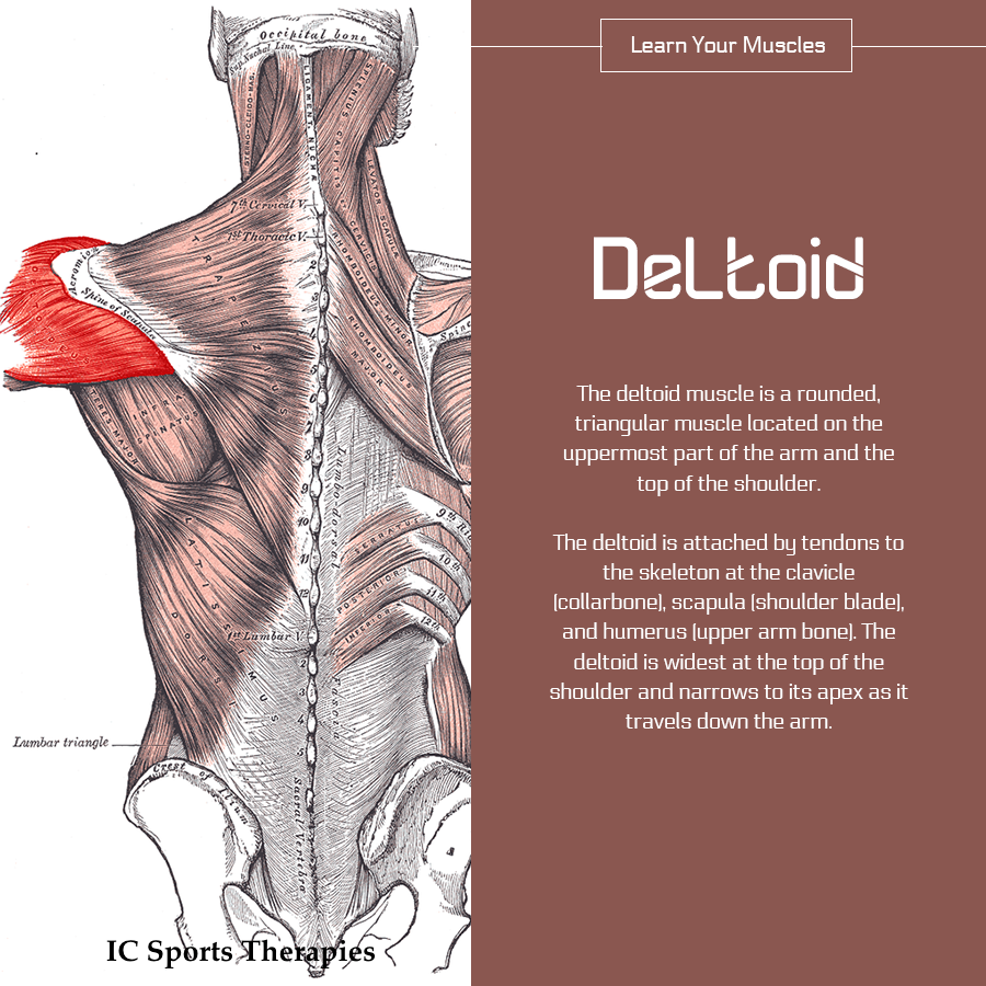
The deltoid muscle is a rounded, triangular muscle located on the uppermost part of the arm and the top of the shoulder. It is named after the Greek letter delta, which is shaped like an equilateral triangle.

The deltoid is attached by tendons to the skeleton at the clavicle (collarbone), scapula (shoulder blade), and humerus (upper arm bone). The deltoid is widest at the top of the shoulder and narrows to its apex as it travels down the arm.

Contraction of the deltoid muscle results in a wide range of movement of the arm at the shoulder due to its location and the wide separation of its muscle fibres… (Read more at InnerBody.com)

The deltoid muscle has three parts; the front or anterior, middle, and back or posterior.

The muscle lifts the arm up sideways. The front part helps to lift the arm up forwards, known as shoulder flexion and the back part helps to lift the arm up backwards, known as shoulder extension.


Previous: Your Monday Muscle: #1 the Trapezius

Next: Your Monday Muscle: #3 Pectoralis Major

Wednesday Wonder #9: Coughs and Sneezes

The cough

The average human cough would fill about three-quarters of a two-liter soft-drink bottle with air — air that shoots out of the lungs in a jet several feet long. Coughs also force out thousands of tiny droplets of saliva. About 3,000 droplets are expelled in a single cough, and some of them fly out of the mouth at speeds of up to 50 miles per hour.

The sneeze

Sneezing is even worse!

It starts at the back of the throat and produces even more droplets — as many as 40,000 — some of which rocket out at speeds greater than 200 miles per hour. The vast majority of the droplets are less than 100 microns across — the width of a human hair. Many of them are so tiny that they cannot be seen with the naked eye.

Airborne and ready to infect

Once airborne, viruses in these tiny droplets can survive for hours. Even if the droplets hit a surface, the viruses can survive and still spread disease if the droplets become airborne later. When a droplet lands on paper, its virus particles can survive for hours. On steel or plastic they can survive for days.

Read more in “The Gross Science of a Cough and a Sneeze


Previous: Wednesday Wonder #8: Smelly Feet

Next: Wednesday Wonder #10: 200 million litres of blood

Your Monday Muscle: #1 the Trapezius

Trapezius

The trapezius

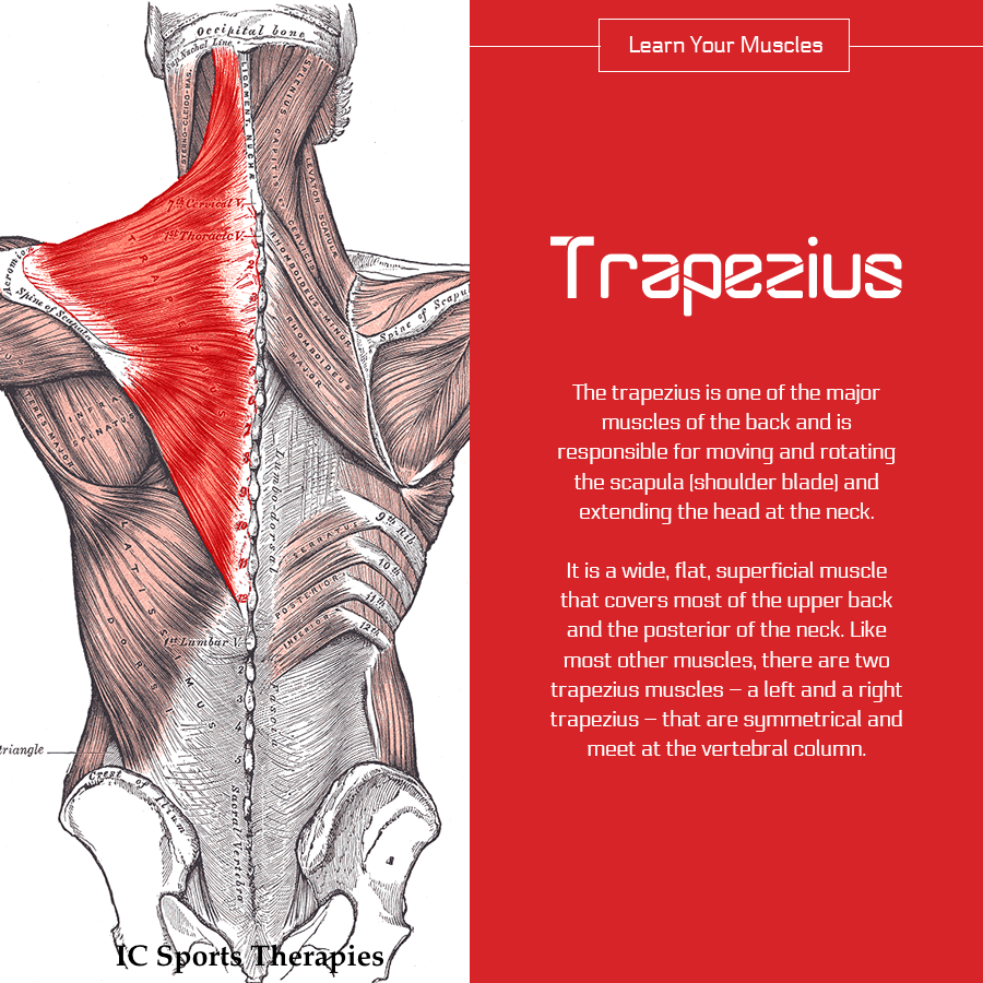
The trapezius is one of the major muscles of the back and is responsible for moving, rotating, and stabilizing the scapula (shoulder blade) and extending the head at the neck. It is a wide, flat, superficial muscle that covers most of the upper back and the back of the neck. Like most other muscles, there are two trapezius muscles – a left and a right trapezius – that are symmetrical and meet at the vertebral column

The trapezius starts at the occipital bone (at the back of the head) and the spinous processes of the cervical and thoracic vertebrae, then extends across the neck and back to insert via tendons on the clavicle (collar bone), acromion (the summit of the shoulder), and spine of the scapula. The name trapezius is given to this muscle due to its roughly trapezoidal shape. The trapezius can be divided into three bands of muscle fibers that have distinct structures and functions within the muscle: Upper Trapezius, Middle Trapezius and Lower Trapezius.

Read more at InnerBody.com

Your Monday Muscle: #2 the Deltoid

Wednesday Wonder #6: the small intestine

This Week’s Wednesday Wonder:

It’s bizarre to think that an organ measuring four times your own height is all coiled up inside your torso, but the small intestine plays a pivotal role in the body. It’s the MVP of your digestive system. (Don’t tell the stomach or colon we said that.) The small intestine—also known as the small bowel—is made up of three parts, which together stretch about 22 feet (6.7 meters) long. Those three parts are the duodenum, the jejunum, and the ileum. (Read more…)

Next: #7 Skin